The difference between a good implant and a great one is often a millimeter of angle, depth or distance from a nerve. Digital implant surgery exists to control that millimeter. Seoul Top Dental Hospital in Seocho explains how the digital workflow runs from scan to surgery, and what it changes for the patient.
One virtual patient: merging CT and oral scan
The workflow starts by combining two data sets: a cone-beam CT showing bone, nerves and sinus, and an intraoral scan capturing teeth and gum surfaces precisely. Software merges them into one virtual model, where the final crown is designed first and the implant position is planned backward from it. This is prosthetically driven planning, not “wherever the bone allows.”
The surgical guide: plan transferred to the mouth
From the approved plan, a custom surgical guide is 3D-printed. Seated on the teeth during surgery, its metal sleeves physically constrain the drill to the planned angle and depth. This narrows the human-error window, often allows smaller incisions and sometimes flapless surgery, and shortens chair time, which patients feel as less swelling afterward.

Who benefits most
Guided surgery shines where margins are tight: implants near the nerve canal or sinus, multiple adjacent implants that must be parallel, immediate placements and full-arch cases where prosthetic alignment is everything. For simple single implants in abundant bone, freehand by an experienced surgeon remains perfectly sound; the diagnosis determines whether the guide earns its place.
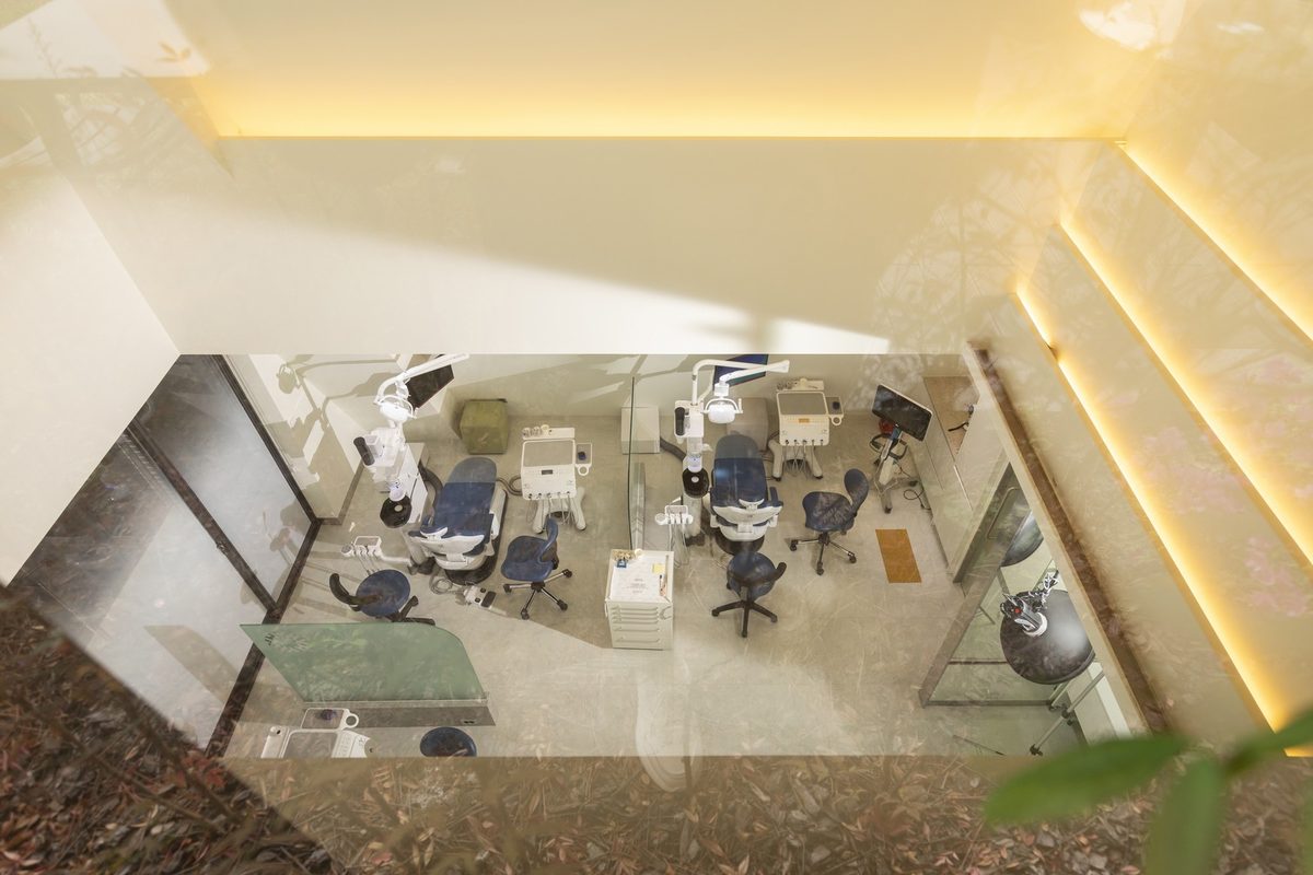
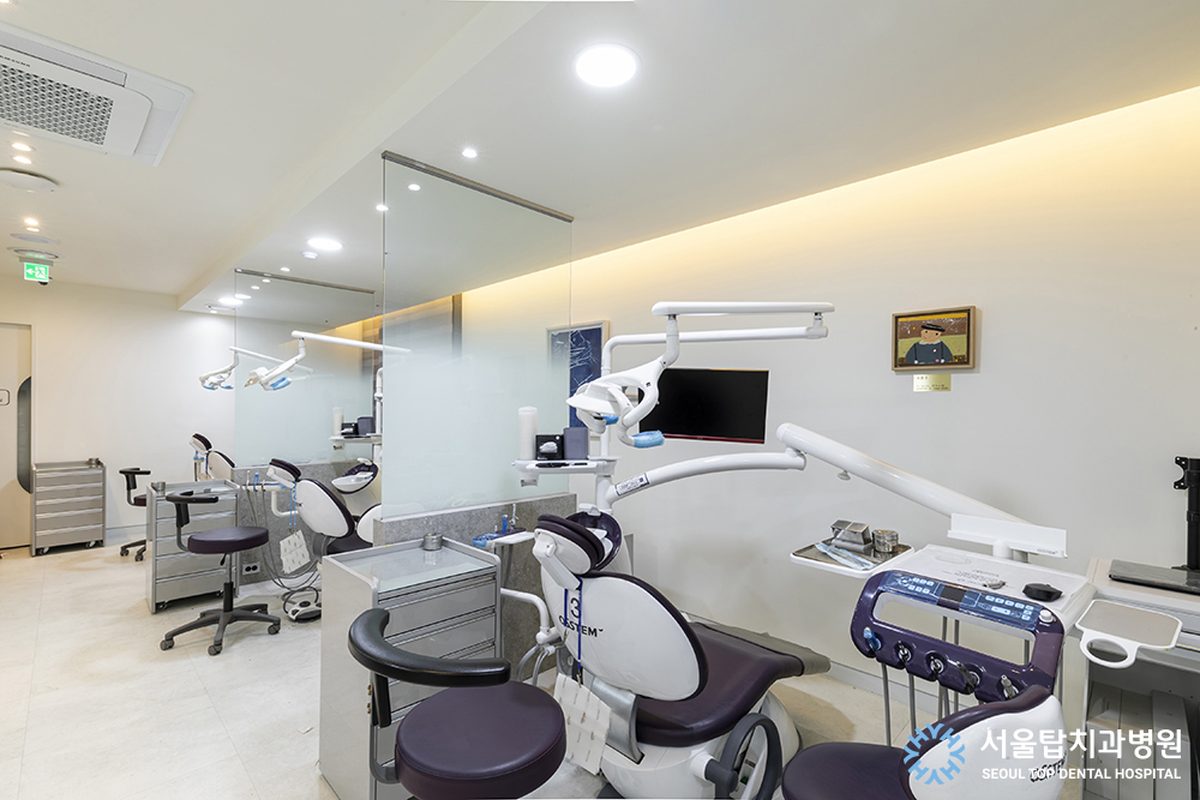
Seoul Top Dental Hospital runs a fully digital planning workflow with in-house CT and scanning, and explains your own 3D plan on screen before surgery. Seeing your implant’s planned position yourself is the clearest way to understand the treatment, so ask for it at consultation.